Describe the Structure of the Wall of the Urinary Bladder
Sensations from the bladder are transmitted to the central nervous system CNS via general visceral afferent fibers GVA. In the epithelium four different cell types were recognized on the basis of their fine structure and staining properties with several different dyes.

Urine Transport And Other Structures Of The Urinary System Anatomy And Physiology
Beneath the urothelium there is a dense capillary plexus.

. Men and women have the same upper urinary tract but their lower urinary tracts are different. Submucosa Lamina Propria of the Bladder Wall The submucosa of the urothelium consists of connective tissue collagen elastin and other extracellular matrix proteins. Pages 21 Ratings 67 3 2 out of 3 people found this document helpful.
1 Ready Set Go 2 Where Why And What 3 Meat And Bones 4 Head To Toe and All Parts In Between 5 What Is In A Name 6 Gut Instincts 7 Null And Void 8 Have A Heart 9 A Breath Of Fresh Air 10 Skin Deep 11 The Great. An Illustrated Guide To Vet Med Term. Muscular wall is expandable and contractible.
Peristalitic waves convey urine along the tube. School University of Phoenix. Muscular tube that conveys urine form pelvis of the kidney to urinary bladder 2.
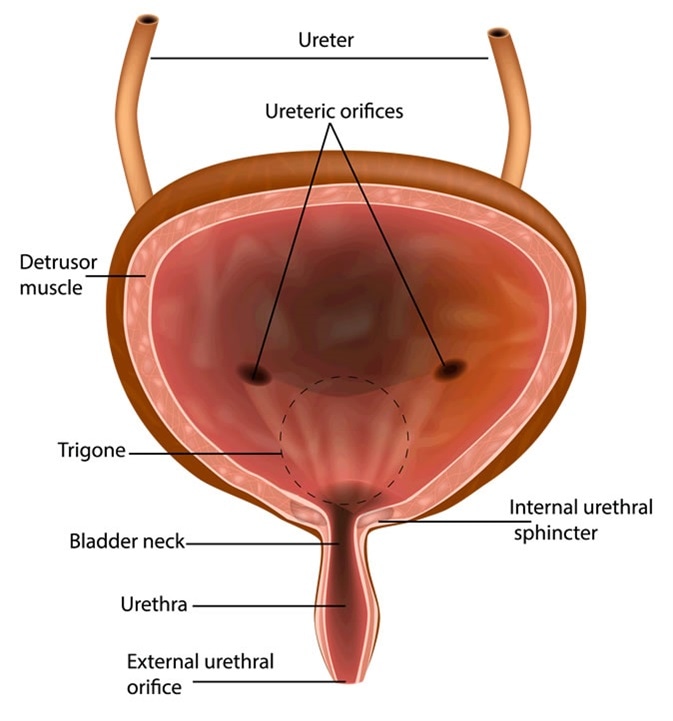
It plays two main roles. The detrusor muscle is the muscular layer of the wall made of smooth muscle fibers arranged in spiral longitudinal and circular bundles. Textbook solution for BIO 2340 ANATOMY-INCLUSIVE ACCESS PT1 15th Edition SHIER Chapter 20 Problem 29P.
The structure of the bladder consists of three main parts. It has a folded internal lining known as rugae which allows it to accommodate up. Structure - distensible organ that is hollow and muscular.
Bladder wall hypertrophy and increased bladder weight are the physiologic responses of the urinary bladder to BOOBPO. Loose Leaf Version of Holes Essentials of Human Anatomy amp. Layers of the Bladder Wall.
Describe the structure of the wall of the urinary bladder. The bladder is an organ of the urinary system. Together the two kidneys and two ureters make up the upper urinary tract.
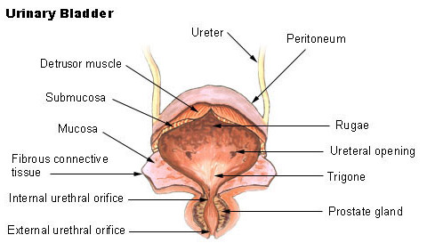
URETHRA YELLOW The urethra is located on either side of your body just above your belly button. The walls of the bladder have a series of ridges thick mucosal folds known as rugae that allow for the expansion of the bladder. The longer BOOBPO exists the thicker the bladder wall and heavier the bladder become in both animals and humans.
Course Title NSCI 281. The urethra which leads from the kidneys to the bladder. Temporary storage of urine - the bladder is a hollow organ with distensible walls.
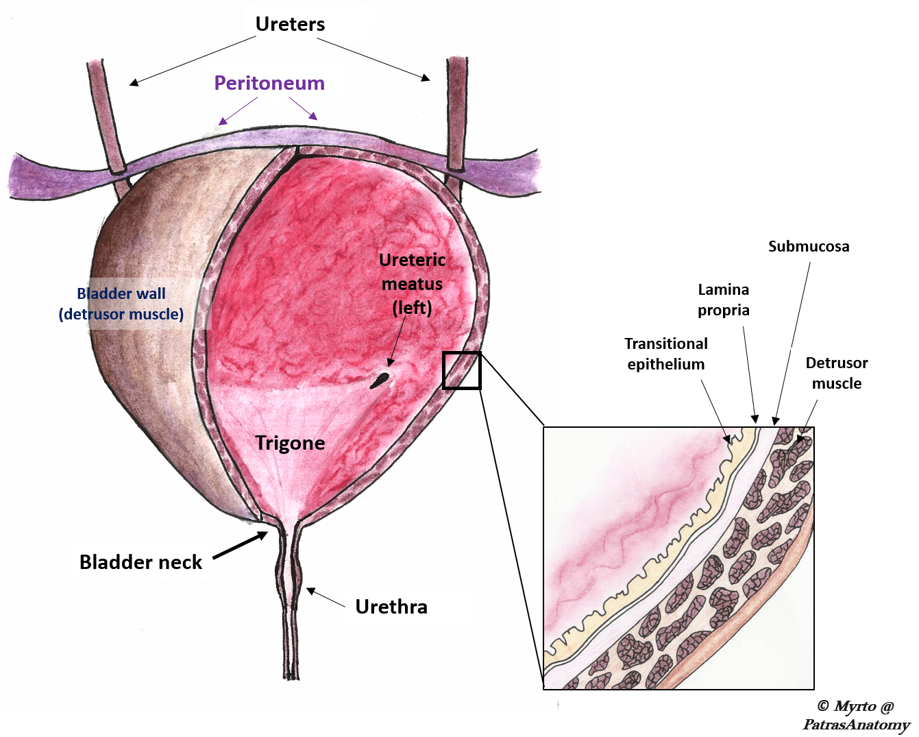
Answer to Describe the structure of the bladder wall. The wall of the urinary bladder has four layers. Lines the bladder ureters and urethra.
This signal will encourage the bladder to expel urine through the urethra. The urinary bladder of the toad Bufo marinus was studied with both the light and the electron microscopeThe bladder wall consists of epithelium submucosa and serosa. The upper urinary tract and the lower urinary tract.
Describe the structure of the urinary bladder hollow. We have step-by-step solutions for your textbooks written by Bartleby experts. The detrusor muscle is a layer of the bladder wall made of smooth muscle fibers that are arranged in spiral longitudinal and circular bundles.
Lining epithelium Lamina propria Muscularis propria SerosaAdventitia Lining epitheliumThe urinary bladder lining is a specialized stratified epithelium the urothelium. The detrusor muscle is able to change its length. Describe the structure function of the urinary bladder The urinary bladder from BIOL 118 at University of Washington.
And the muscular wall that surrounds these two organs. The wall of the bladder wall has three principal tissue layers or coats. Location - within the pelvic cavity posterior to the symphasis pubis and inferior to the parietal peritoneum.
This acts as a lining. In addition to the vascular supply of the urothelium the capillary plexus serves as a barrier function. The outside layer is either serosa or adventitia depending on location--see your textbook for an explanation.
The detrusor muscle contracts around the ureteric orifices when the bladder contracts in order to prevent vesicoureteral reflux backflow of urine into the ureters. It forms the internal urethral sphincter around the neck of the bladder. The lower urinary tract contains the bladder and the urethra.
From the inside towards the outside they are. The detrusor muscle comprises the wall of the urinary bladder. Answer to Describe the structure of the bladder wall.
Features and Structure of the Bladder The transitional epithelium layer is the first layer on the inside of the bladder. Muscular wall of the urinary bladder that surrounds the neck of the bladder and forms an internal urethral sphincter. Describe the structure of the urinary bladder Hollow distensible muscular organ.
The microscopic structure of the urinary bladder wall organizes into the following layers from inside out. Mucosa submucosa muscularis and serosa or adventitia. Connect Plus LearnSmart 2 Semester Access Card for Holes Human Anatomy and Physiology 13th Edition Edit edition.
What is the medical term meaning hernia of. Initial investigations focused on the ultrasound appearance of the bladder wall and demonstrated that the detrusor appears as a. The urinary system has two parts.
The urinary sphincter which separates urine from other wastes. Mucous membrane mucosa Transitional epithelium.

The Urinary Bladder Structure Function Nerves Teachmeanatomy


Comments
Post a Comment